


Rehab Medical Workshop


Doppler Ultrasound Machines Main Functions
Having a full digital beam forming technology
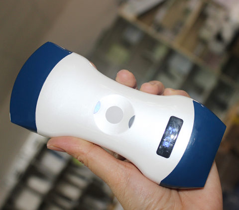
Scanning mode: Convex array, lumen, high-frequency linear array, phased array, 3D software (Optional accessories);
Dynamic range: 0~120dB adjustable;
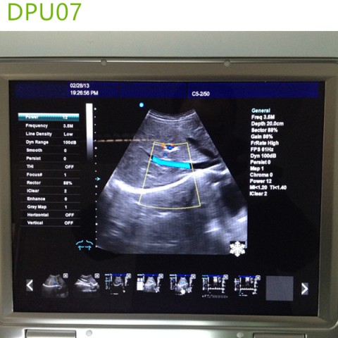
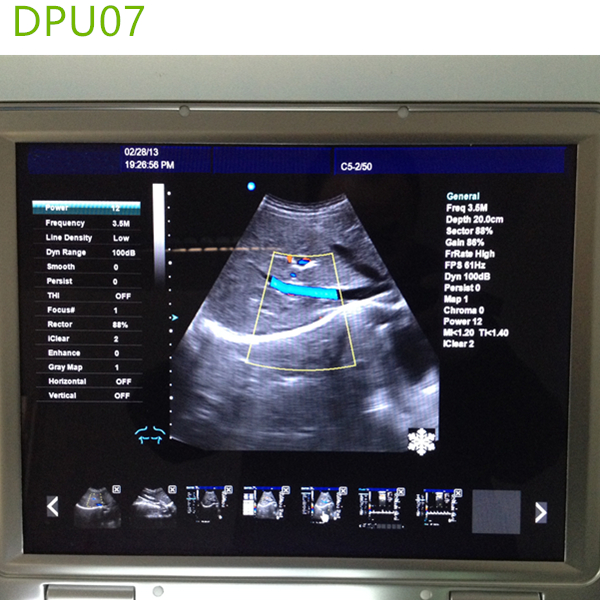
Display mode: B、B/B、M、B/M、CFM、CMF/B、PDI、B/PW, total eight mode;
Application mode: abdomen, gynecology, obstetrics, superficial organ, urologist, heart and user defined model 1-4, total ten models;
Image mode: digital beam forming, tissue harmonic imaging;
Acoustic output: Mechanical index and thermal index real-time display;
Acoustic power:Step is adjustable, real-time display;
Gray scale: 256 scales;
Depth display: ≥250mm;
B/D dual-purpose: linear array: B/PWD; convex array: B/PWD;
Pseudo color processing: 16 kinds of pseudo color encoding can optional;
Gain adjusts: 8 segments TGC, B/M/D/C is independently adjustable; TGC curve can show and hide automatically;
Image magnification: picture in picture zoom in and zoom part function;
Image processing: Edge enhancement: Multilevel adjustable
Frame average: Multilevel adjustable
Line average: Multilevel adjustable
Focus Optimization: Multilevel adjustable
Gray Restrain: Multilevel adjustable
Gamma correction: Multilevel adjustable
Contrast: Adjustable
Brightness: Adjustable
Self-motion optimize function: Built-in multiple check type, according to different inspection organs, preset best image check condition, reduce the adjusting operation keys;
One-click optimization function: preset several parameters adjusting focus on a button, a key to realize image fast optimization;
Measurement and calculation: B mode routine measurement:
Distance, circumference, area, volume, angle, ratio, and stenos rate.
M mode routine measurement: Heart rate, time, distance, speed, ratio, etc..
Gynecology measurement: Uterus, cervix, endometrial, ovary, follicular.
Obstetrics measurement:
EGA, ETD, fetal weight estimation, AFI index, OB report (including OB tables).
Cardiology measurement: LV measurement.
Urology measurement:
Prostate volume, displacement volume, bladder capacity, and residual urine output.
PW measurements: Time, speed, Heart Rate, RI, PI, etc.
Other measurement: Slice volume measurement, hip joint angle measurement.
Image storage: Image storage, video storage, cine loop, disk storage capacity≥160G;
Patient data: Medical record management, report inquiry and printing, image video output( HDD 、 USB)、built-in ultrasound workstation;
Reporting system: automatic report generation system, and can be full screen characters in both Chinese and English editor;
Output interface: SR323、USB、DICOM interface;
Laptop Doppler Ultrasound Main Technical Indexes
The performance requirements of gray-scale imaging mode
The color ultrasonic at the gray-scale imaging performance mode should comply with the provisions of the table 2.1
Table 2.1 At the Gray-scale imaging mode the performance of the probe
| performance indexes | probe type and nominal frequency | |||
| 2.0≤f<4.0 | 2.0≤f<5.0 | 5.0≤f<8.0 | 5.0≤f<10.0 | |
| a) probe type and model | phased array (type TP16A) | Convex array
(type TC60A) |
Cavity
(type TC10A) |
Linear array
(type TL40A) |
| b) nominal frequency (MHz) | 3.0 | 3.5 | 6.5 | 7.5 |
| c) Scan depth(mm) | ≥140 | ≥160 | ≥40 | ≥50 |
| d) Lateral resolution (mm) | ≤3(depth≤80)
≤4(80<depth≤130) |
≤3(depth≤80)
≤4(80<depth≤130) |
≤2(depth≤30) | ≤2(depth≤40) |
| e) Axial resolution (mm) | ≤2(depth≤80) | ≤2(depth≤80)
≤3(80<depth≤130) |
≤1(depth≤40) | ≤1(depth≤50) |
| f) Blind area (mm) | ≤7 | ≤5 | ≤4 | ≤3 |
| g) Transverse geometry precision (%) | ≤20 | ≤15 | ≤10 | ≤10 |
| h) Longitudinal geometric location accuracy (%) | ≤10 | ≤10 | ≤5 | ≤5 |
| i) Slice thickness (mm) | ≤5 | ≤5 | ≤5 | ≤5 |
| j) Perimeter and area measured deviation (%) | ≤±20 | ≤±20 | ≤±20 | ≤±20 |
| k) M mode time display error (%) | ≤±10 | ≤±10 | ≤±10 | ≤±10 |
The performance requirements of color Doppler imaging mode
a). The color ultrasonic at the color Doppler imaging mode should comply with the provisions of the
table 2.2;
b). Color blood flow image should be essentially coincident with the gray-scale image of pipe’s;
c). Blood flow direction should be able to correctly identify, no aliasing phenomenon;
Table 2.2 at the color blood flow imaging mode the performance of the probe
| Doppler model | Phased array | Convex array | Cavity | Linear array |
| Investigation depth at Color blood flow model | ≥90mm | ≥100mm | ≥40mm | ≥50mm |
| Investigation depth at Doppler spectrum model | ≥90mm | ≥100mm | ≥40mm | ≥50mm |
| Blood flow speed reading error | ≤±15% | |||
The performance requirements of Doppler spectrum mode
a). The color ultrasonic at the color Doppler spectrum mode should comply with the provisions of the;
b). Blood flow speed reading error should comply with the provisions of the table2.3;
c). Pulse wave Doppler mode sampling area cursor position should be accurate;
Table 2.3 at the color blood flow imaging mode the performance of the probe
| Doppler model | Phased array | Convex array | Cavity | Linear array |
| Investigation depth at Color blood flow model | ≥90mm | ≥100mm | ≥40mm | ≥50mm |
| Investigation depth at Doppler spectrum model | ≥90mm | ≥100mm | ≥40mm | ≥50mm |
| Blood flow speed reading error | ≤±15% | |||
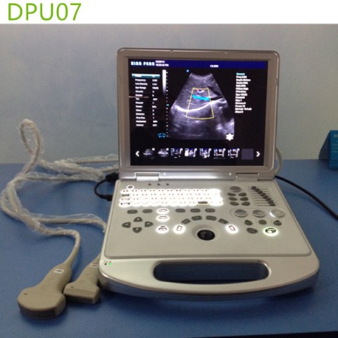
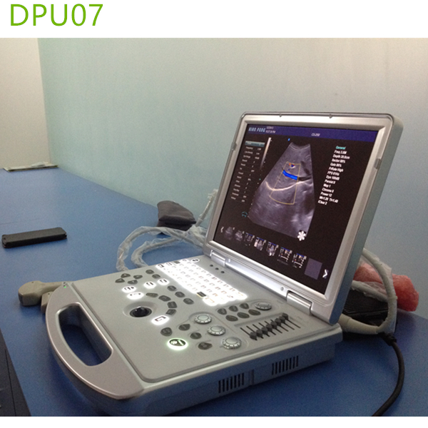
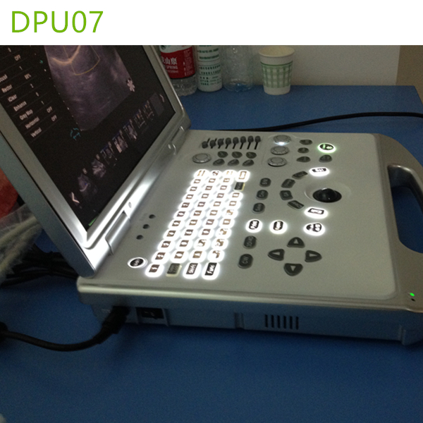
Display: 15 inch LED color display
Running hours: ≥8h;
Input power: ≤320V;
Host weight: about 9.5 kg;
Battery life:2 hours
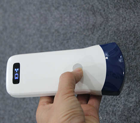
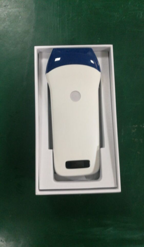
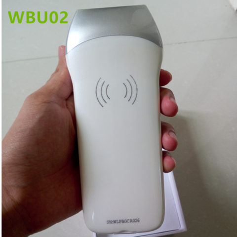
More pictures about hot sale portable doppler ultrasound machines
 Packaging detail:Standard export package.
Delivery detail:within 7-10 working days after receipt of payment.
Warranty Period :12-24 months (stats from the day arrival destination port)